A review of Developmental Dysplasia of the Hip (DDH).
DDH occurs due to instability from ligamentous laxity and intrauterine/postnatal malpositioning. Instability leads to dysplasia which may lead to subluxation and dislocation.
It is more common in females, firstborns, and breech births.
Barlow (dislocation), Ortolani (relocation), and Galeazzi (limb length) testing may be used in infants < 3 m.o.
In children, 3 mo. to 1 yr. of age, leg length discrepancy, limited abduction, and positive Klisics test (shown below) may indicate DDH.

In children > 1 year of age pelvic obliquity, lumbar hyperlordosis, Trendelenburg gait (due to limited abduction), and toe walking (compensation for leg length discrepancy) may be seen.
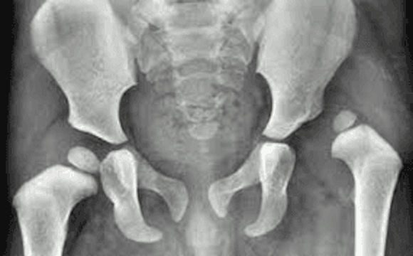
Children less than four months of age should undergo ultrasound imaging for suspected DDH. Between 4-6 months of age, radiography becomes the imaging modality of choice.
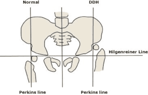
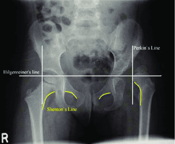
Measurements are performed using an AP Pelvis.13
Measurements to screen for hip dislocation: Hilgenreiner's line, Perkin's Line, and Shenton's Line.
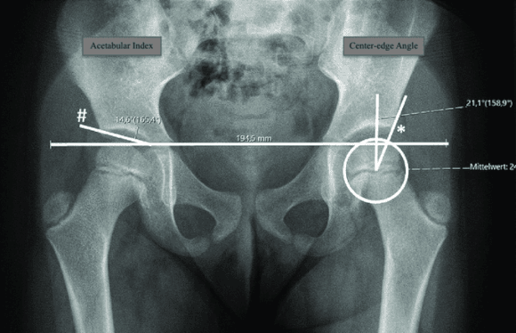
Measurement to screen for hip dysplasia: acetabular index and central-edge angle.
Hilgenreiner's line is a horizontal line through the triradiate cartilages. Femoral head ossification(FHO) should lie inferior to this line.
Perkin's line is drawn perpendicular to the Hilgenreiners line at the lateral aspect of the acetabulum, FHO should lie medial to this line
Shenton's line is drawn along the inferomedial femoral neck and inferior border of the superior pubic rami. The line should form a continuous arc.
The acetabular index is formed by Hilgenreiner's line and a line formed between the lateral triradiate cartilage and lateral acetabulum.
This line should be < 25° in children > 6 m.o. old.

The center-edge angle (Wiberg angle) is formed by a line perpendicular to Hilgenreiner's line (Perkins line) and a line between the center of the femoral head and the lateral edge of the acetabulum.
Used in children > 5 y.o.
< 20° is abnormal.
Non-operative treatment options include Pavlik harness and closed reduction with spica casting.
Operative treatment options include open reduction with spica casting, OR with femoral osteotomy, and OR with pelvic osteotomy.
Full Review and Conversation on X